On radiographic images, what feature is often what ultimately makes an established Romanus lesion definitively identifiable?
Answer
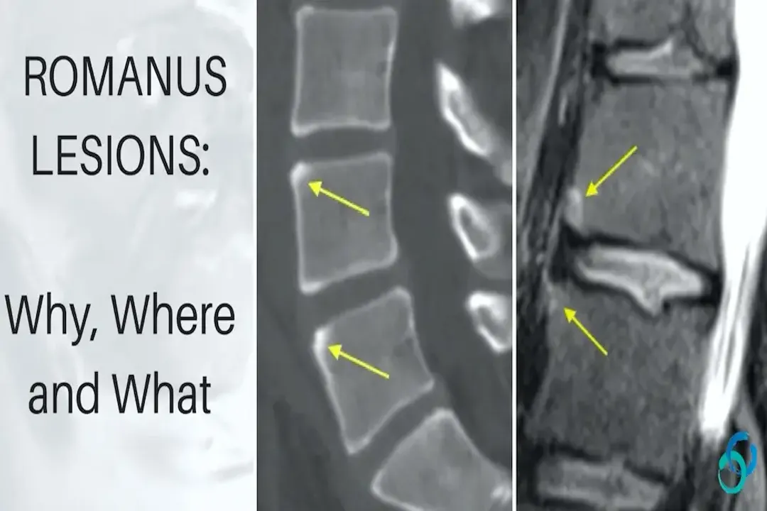
Increased density (sclerosis)
While the initial event of a Romanus lesion is erosive damage visible as small defects, the feature that typically renders the lesion clearly identifiable on standard X-rays or CT scans is the subsequent reparative process. Following erosion, the body attempts to heal by depositing new, dense bone tissue, resulting in reactive sclerosis. It is this sclerosis, appearing as a pathologically dense, whiter area adjacent to the disc space at the vertebral margins, that provides the definitive radiographic signature of an established Romanus lesion, often obscuring the initial subtle erosion.
Related Questions
What specific inflammatory process drives the bony changes resulting in a Romanus lesion?What initial destructive effect on the bone characterizes the nascent stage of a Romanus lesion?What established lesion results from the maturation and extensive repair of an initial Romanus lesion?In which spinal regions are Romanus lesions most frequently identified radiologically?What early finding detectable by MRI reflects the ongoing inflammation in an active Romanus lesion?A key feature distinguishing a Romanus lesion is its dual nature showing both damage and what reparative sign?How does the primary destruction pattern in typical vertebral osteomyelitis contrast with that of a Romanus lesion?The enthesitis causing Romanus lesions specifically targets the insertion point of which structure onto the vertebral body?Identifying a Romanus lesion signals the initiation of the pathological process leading toward what advanced AS spinal feature?On radiographic images, what feature is often what ultimately makes an established Romanus lesion definitively identifiable?