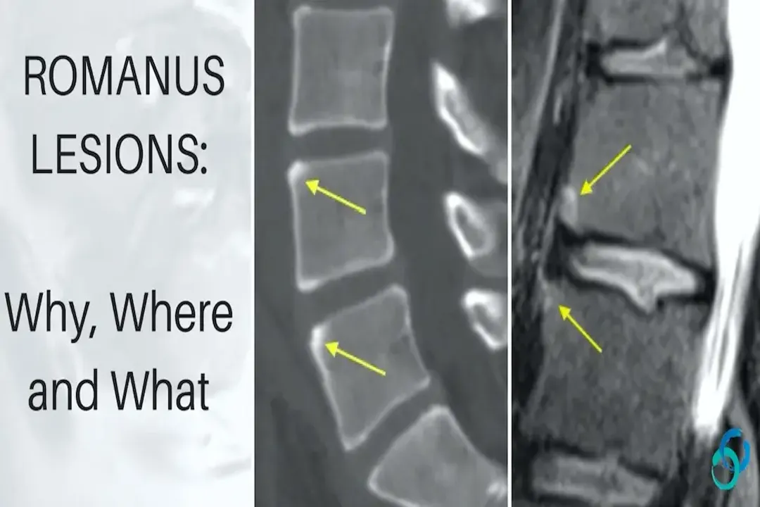
In which spinal regions are Romanus lesions most frequently identified radiologically?
Answer
Lower thoracic and upper lumbar spine
Radiological examination of patients with Ankylosing Spondylitis often reveals a predictable distribution for spinal inflammatory changes. Romanus lesions specifically show a predilection for the areas experiencing significant biomechanical load coupled with inflammatory insult within the axial skeleton. The text specifies that these lesions are most frequently found located within the lower thoracic spine and extending into the upper lumbar spine segments.
Related Questions
What specific inflammatory process drives the bony changes resulting in a Romanus lesion?What initial destructive effect on the bone characterizes the nascent stage of a Romanus lesion?What established lesion results from the maturation and extensive repair of an initial Romanus lesion?In which spinal regions are Romanus lesions most frequently identified radiologically?What early finding detectable by MRI reflects the ongoing inflammation in an active Romanus lesion?A key feature distinguishing a Romanus lesion is its dual nature showing both damage and what reparative sign?How does the primary destruction pattern in typical vertebral osteomyelitis contrast with that of a Romanus lesion?The enthesitis causing Romanus lesions specifically targets the insertion point of which structure onto the vertebral body?Identifying a Romanus lesion signals the initiation of the pathological process leading toward what advanced AS spinal feature?On radiographic images, what feature is often what ultimately makes an established Romanus lesion definitively identifiable?