What is the role of MD in drug discovery?
When we talk about bringing a new medicine from the lab bench to a patient, the process is notoriously long, expensive, and filled with failure points. At the heart of understanding how a potential drug works—its interaction with the body's machinery—lies computational chemistry, and within that field, Molecular Dynamics (MD) simulations play an increasingly vital part. [1][9] MD is essentially a sophisticated virtual laboratory that allows scientists to observe the movements and behaviors of atoms and molecules over time, providing a dynamic view that static pictures simply cannot capture. [1][7]
# Simulation Basics
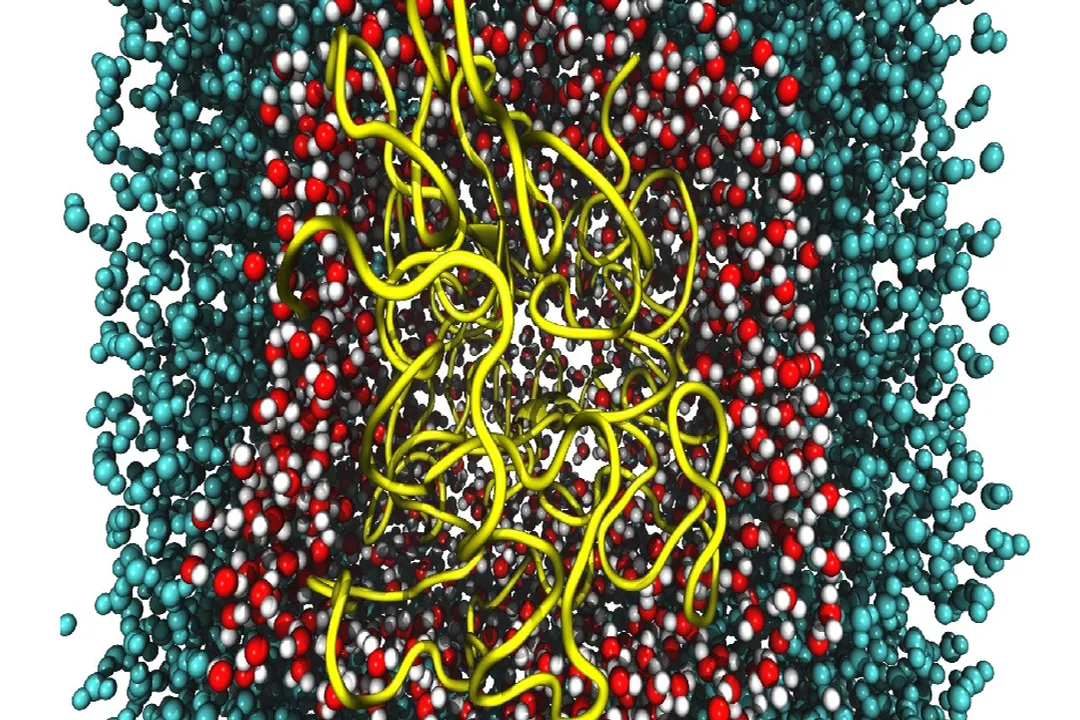
Molecular Dynamics is a computational technique that models the physical movements of atoms and molecules, essentially solving Newton's laws of motion for every particle in the system. [1][9] Instead of providing a single, frozen snapshot of a drug bound to its target protein, MD generates a movie. [1] This movie is built by calculating the forces acting on each atom based on their current positions and then calculating how much each atom will move over a very small time step, often measured in femtoseconds (one quadrillionth of a second). [7] By stringing together millions of these tiny steps, researchers can simulate timescales ranging from nanoseconds to microseconds or even milliseconds. [2][7]
This ability to observe motion is critical because biological systems are inherently dynamic. A protein target, like an enzyme involved in a disease pathway, is not a rigid lock; it breathes, shifts its shape, and opens or closes binding pockets. [1] A small molecule drug candidate must successfully navigate this dynamic landscape to exert its effect. [4]
# Target Understanding
Before a company even begins synthesizing large libraries of compounds, they need to know if their chosen biological target is "druggable." This is where MD simulation enters the early stages, often associated with target validation. [4]
MD can help determine the structural stability of the target protein itself. [1] If the protein, in the environment that mimics the body (often involving explicit water molecules and ions), is prone to unfolding or significant structural collapse under certain physiological conditions, it might be a poor drug target, regardless of how well a potential drug fits its initial crystal structure. [7]
Furthermore, MD simulations allow researchers to look closely at the actual binding site—the groove or pocket where the drug is supposed to dock. [4] While standard docking programs provide a static prediction of the best fit, MD simulates the process of binding. For instance, a drug might look perfect in a static binding pocket, but MD might reveal that the pocket closes too quickly, or that the drug induces a conformation that makes binding energetically unfavorable. [4] This detailed look at the environment surrounding the target provides crucial context that purely static structure analysis misses. [1]

# Hit Refinement
Once initial "hits"—compounds that show weak activity against the target—are identified, the focus shifts to lead optimization, a phase where the goal is to improve potency and drug-like properties. [4][5] MD simulations are incredibly powerful here because they can estimate the binding free energy of a drug candidate to its target. [7]
Binding free energy calculation essentially quantifies how strongly the drug will stay bound under physiological conditions, which correlates directly with how effective the drug will be at lower concentrations. [7] Simpler methods like traditional docking give a score, but MD-based methods, like rigorous free energy perturbation or thermodynamic integration, offer a more physically grounded prediction of this energy. [7]
Consider this scenario: Two compounds, Compound A and Compound B, both dock perfectly into the active site, achieving similar initial scores. If Compound A forms a few critical hydrogen bonds that are maintained consistently throughout a 100-nanosecond MD simulation, while Compound B’s critical interactions are lost due to subtle protein movement every 50 nanoseconds, the MD results strongly suggest Compound A will be the superior lead. [4] This kind of comparative analysis saves significant resources by prioritizing the chemically stronger candidate before it even enters expensive biological assays. [8]
| Simulation Metric | Information Provided | Impact on Lead Optimization |
|---|---|---|
| RMSD/RMSF | Stability of the complex over time; flexibility of regions | Identifies structural collapse or unwanted ligand flapping [1] |
| Binding Free Energy | Quantitative measure of binding strength | Ranks candidates based on thermodynamic favorability [7] |
| Water/Ion Movement | Role of surrounding solvent in stabilization | Suggests ways to modify the drug to displace unfavorable water molecules [1] |
# Computational Speed Comparison
It is helpful to view MD as occupying a specific niche between very fast, low-resolution screening methods and slow, high-resolution experimental techniques. [2][8] High-throughput screening (HTS) checks millions of compounds quickly, but often yields many false positives or compounds that bind poorly in a complex environment. [8] Standard docking is faster than MD but lacks dynamic insight. [4] MD simulations, by contrast, require significant computational power and time, meaning they cannot screen millions of molecules. [2]
The value proposition here is quality over quantity. [8] Instead of screening a million compounds statically, researchers might use MD to rigorously test the top few hundred candidates suggested by earlier, faster computational methods. [5] This selective, in-depth computational validation acts as a very effective filter, ensuring that only the most promising, dynamically stable compounds move forward to resource-intensive laboratory testing. [8] The investment of time in an MD simulation yields a much higher confidence level in the compound's viability than a simple static fit calculation.

# Complement to Experiment
MD simulations are rarely intended to replace experimental validation like X-ray crystallography or Nuclear Magnetic Resonance (NMR) spectroscopy; rather, they are designed to complement and interpret these methods. [2][3]
When an experimental structure is solved—perhaps showing a drug bound to a protein—it represents a single, low-energy state that the system adopted long enough to be captured. [1] MD can take that static picture and explore the pathway the system took to get there, or, more importantly, explore other relevant, near-minimum energy states that the experiment might have missed. [1][3] For example, NMR or crystallography might show a ligand deep in a pocket, but MD simulations can reveal that the pocket must temporarily widen by 2 Angstroms, requiring a specific degree of flexibility from the protein residues, information essential for medicinal chemists designing analogs. [2]
One of the key contributions of MD to structural biology efforts, as noted in research, is the ability to model the impact of mutations or post-translational modifications on ligand binding, which can be directly tested or compared against experimental mutagenesis studies. [6] When experimental data shows a compound doesn't work as expected after a mutation, MD provides the atomic-level hypothesis for why—perhaps the mutation destabilized a key loop necessary for the drug's proper orientation. [6]
# Practical Application Example
Imagine a team designing a kinase inhibitor. They have a promising lead, Compound C, which fits well into the ATP-binding site. Initial docking suggests it’s strong. However, the team observes that the protein target possesses a flexible "activation loop" near the binding site. Through a few microseconds of MD simulation, the researchers observe that while Compound C initially binds, the activation loop shifts into a conformation that sterically clashes with a bulky part of Compound C after just 50 nanoseconds, forcing the molecule into a slightly misaligned, less effective pose. [4] A simpler docking score would have been optimistic. This subtle, time-dependent rejection by the simulated protein environment would immediately tell the chemists to go back and redesign the bulky part of Compound C to be smaller, thereby accommodating the necessary loop motion—a costly error avoided entirely in the computer before ever synthesizing the next batch of molecules. [8]

# Future Trajectory
While MD offers incredible detail, its major hurdle remains the timescale barrier. [2][7] Important biological events, like a ligand fully escaping a deep binding pocket or large domain rearrangements, occur on microsecond or even millisecond scales, which are computationally expensive to simulate directly with high accuracy. [2][7] Advances in computational power, specialized hardware (like GPUs), and improved sampling methods (like enhanced sampling techniques) are steadily pushing the reachable timescale forward. [2][7]
The general consensus within computational drug discovery is that MD is moving from a specialized validation tool to a more integrated part of the design cycle, driven by better algorithms and increased access to high-performance computing resources. [5][8] Its role is solidifying as a high-resolution microscope for understanding the mechanism of interaction, ensuring that the molecules designed for tomorrow's therapies are optimized not just for fit, but for true dynamic performance within the complex living environment. [1][9]
Related Questions
#Citations
Role of Molecular Dynamics Simulations in Drug Discovery
Role of MD in Drug Discovery - CECAM
Role of Molecular Dynamics and Related Methods in Drug Discovery
The role of molecular dynamics simulations in drug discovery | Cresset
Molecular Dynamics Simulations in Drug Discovery and ... - MDPI
Structure and dynamics in drug discovery - Nature
Molecular modeling in drug discovery - ScienceDirect.com
How MD simulations helped in drug discovery - LinkedIn
Molecular Dynamics Simulation in Drug Discovery and ...