What is the life expectancy of blue babies?

The term "blue baby" historically refers to an infant born with severe cyanosis, a condition where the baby's skin, lips, and nail beds appear bluish or purplish due to a lack of oxygenated blood circulating in the body. This startling visual sign is almost always the result of a serious congenital heart defect present at birth. While various defects can cause this, the most common and historically significant condition associated with the "blue baby" moniker is Tetralogy of Fallot (TOF). Understanding the life expectancy for these children requires looking at the condition through a historical lens, tracing the dramatic changes brought about by decades of advancements in pediatric cardiology and cardiac surgery.
# Cyanosis Mechanism
The bluish discoloration itself is the outward manifestation of hypoxemia, or low oxygen levels in the arterial blood. In a healthy heart, oxygen-poor blood returning from the body goes to the lungs to pick up oxygen before being pumped out to the rest of the body. In a "blue baby," a structural problem in the heart allows this deoxygenated blood to shunt directly into the systemic circulation—the flow meant for the rest of the body—bypassing the lungs. This mixing or shunting means the circulating blood cannot carry enough oxygen, leading to cyanosis. The severity of the blueness often correlates directly with the degree of shunting and the resulting oxygen deprivation, which places significant strain on every organ system.
# Heart Defect Basics
For readers to grasp the prognosis, it is helpful to understand the underlying anatomy of the most common cause, TOF. Tetralogy of Fallot is characterized by four distinct, yet interrelated, structural abnormalities in the heart, all existing simultaneously. These are:
- A large hole between the two lower chambers of the heart, known as a Ventricular Septal Defect (VSD).
- An obstruction to the right ventricle outflow, typically caused by pulmonary stenosis (a narrowing of the pulmonary valve or the area below it).
- The overriding aorta, meaning the large artery that carries blood out to the body is positioned directly over the VSD, allowing it to receive blood from both ventricles (the oxygen-poor blood from the right and oxygen-rich blood from the left).
- Right Ventricular Hypertrophy, where the muscle wall of the right ventricle thickens due to the increased workload caused by the obstruction at the pulmonary valve.
It is the combination of the VSD and the pulmonary stenosis that permits the blood to shunt right-to-left, leading to cyanosis. Other conditions can also present as cyanotic heart disease in infants, but the treatment pathways and eventual outcomes are often compared against the history established by TOF management.

# Early Prognosis
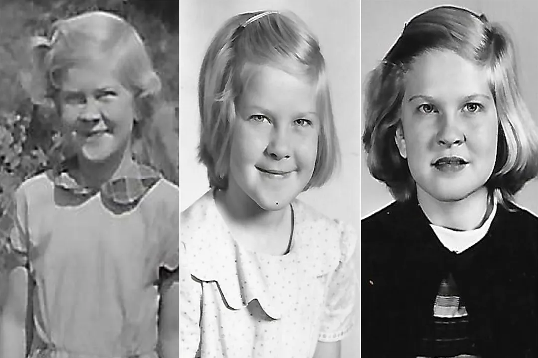
Prior to the advent of modern cardiac surgery, the outlook for infants born with significant cyanotic heart disease like TOF was profoundly poor. Without intervention, many infants would not survive past infancy or early childhood. The chronic lack of oxygen caused developmental delays, poor growth, and frequent, severe episodes known as "tet spells" or "blue spells," where the baby would become intensely cyanotic and often hypoxic, sometimes requiring emergency measures. Survival into adulthood was exceedingly rare; for context, a patient who successfully navigated childhood with uncorrected TOF and lived into their sixties represents a rare success story from an era before definitive surgical correction became routine. Historically, the survival statistics were grim because the only options available were supportive care, which could not fix the underlying plumbing issue.
# Surgical Repair Era
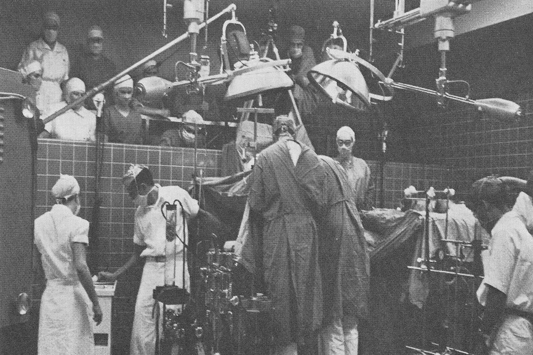
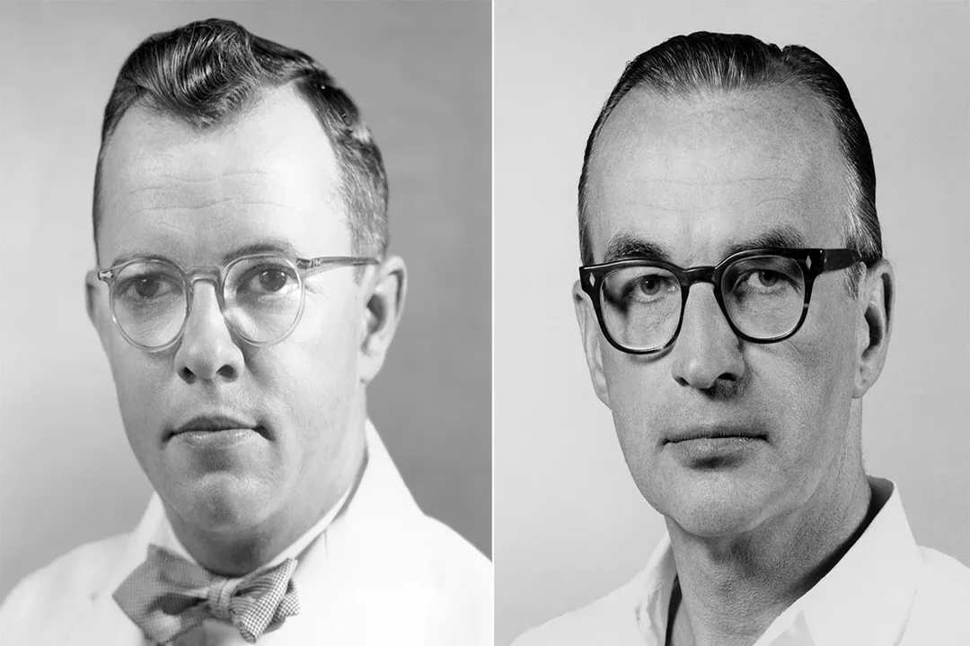
The trajectory of life expectancy shifted dramatically with the introduction of surgical correction, moving the condition from a near-certain death sentence to a treatable defect. Initial surgical attempts in the mid-20th century were often palliative, meaning they aimed to temporarily improve blood flow to the lungs to sustain the child until they were strong enough for full repair. A classic example of a palliative approach was the creation of a shunt, such as a Blalock-Taussig (BT) shunt, which created an artificial connection between a major artery from the body and the pulmonary artery, thereby increasing blood flow to the lungs to oxygenate the blood. This surgical lifeline allowed many children to gain weight, grow stronger, and survive the high-risk neonatal period.
The true turning point came with the development of techniques for complete surgical correction, which allowed surgeons to close the VSD and relieve the obstruction at the pulmonary outflow tract in a single operation. A major study published in The Lancet in 2004, reflecting on patients from earlier decades, highlighted that survival rates for these severe defects were heavily dependent on the era of diagnosis and treatment, underscoring the monumental impact of surgical timing and technique. It is this progression—from palliative shunts to full repair—that forms the foundation of modern life expectancy figures.

# Current Survival Rates
In contemporary medicine, the life expectancy for a child diagnosed with Tetralogy of Fallot, when treated promptly with surgery, is vastly different from historical outcomes. Modern series report excellent survival rates following complete repair in infancy or early childhood. For instance, the survival rate following the first heart surgery for TOF is often cited as being very high, with many series reporting survival rates exceeding 95% for the initial surgery itself.
When considering the overall life expectancy compared to the general, unaffected population, the picture is one of significant improvement but with lingering caveats. Many individuals who underwent successful repair in infancy now live active adult lives. A key piece of evidence comes from tracking these cohorts: patients treated decades ago are now surviving well into their middle and older adult years, suggesting life expectancies are approaching those of their peers without congenital heart disease, though perhaps with a slight reduction depending on the specifics of their defect and repair. One study noted that survival rates for children born with congenital heart defects in the 1980s and 1990s who underwent surgery were significantly better than those born earlier, demonstrating sustained improvement over time.
If we consider the broad spectrum of what constitutes a "blue baby," where some milder forms might only require a single intervention and others may have complex residual anatomy, the average is necessarily broad. However, for the most common severe presentation, TOF, the baseline expectation today is to reach adulthood. It is important to realize that the outcome is rarely "cured" in the absolute sense; rather, the defect is repaired or palliated, requiring lifelong monitoring.
# Life After Repair
While the initial surgery grants survival, the long-term management focuses on the consequences of the initial defect and the surgical correction itself. Even with a technically successful full repair of TOF, the heart has undergone significant stress and structural alteration. The heart muscle, having worked against high pressure for months or years before surgery, may have residual issues. Furthermore, the pulmonary valve, which was likely narrowed (pulmonary stenosis), is often either patched or replaced during the initial repair. Over time, this repaired or replaced valve can develop pulmonary regurgitation (leaking). This regurgitation forces the right ventricle to handle a larger volume of blood returning from the lungs during the resting phase of the heartbeat, potentially leading to right ventricular enlargement over many years.
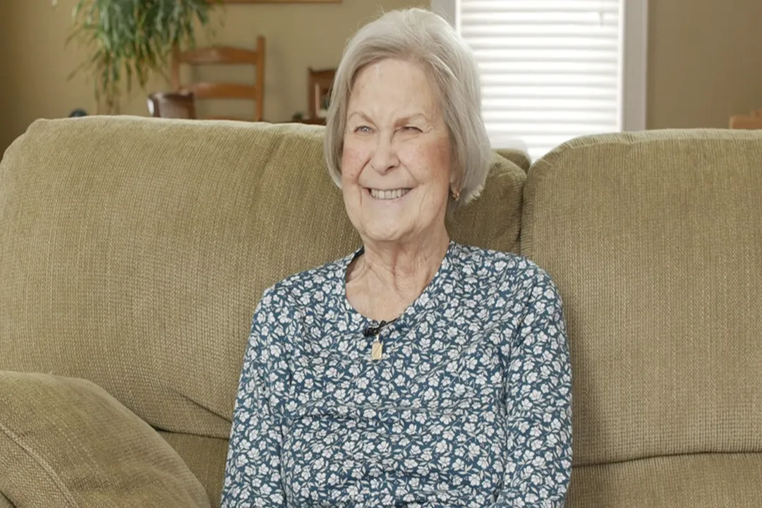
It is this long-term hemodynamic change that dictates the need for continued specialized care. A patient living successfully for decades post-repair, like the individual mentioned who thrived over 65 years, still required specialized attention for their complex, repaired heart. This ongoing management involves regular echocardiograms and other cardiac assessments to monitor for issues like residual leaks, rhythm disturbances, or the need for future valve replacement surgery. In thinking about the lifespan of a "blue baby," the discussion shifts from if they will live to how long their repaired heart structure will function optimally before further intervention is needed.

# Considering Unrepaired Defects
While modern protocols strongly favor early intervention, some historical cases or cases in regions with limited access to specialized care might present with unrepaired or partially repaired defects in older children or adults. For these individuals, the life expectancy is severely truncated compared to those who received timely repair. Chronic, uncorrected cyanosis leads to significant organ damage over time due to persistent hypoxia and polycythemia (an increase in red blood cells to compensate for low oxygen). Survival past early adulthood without correction is rare and typically associated with substantial physical limitations and severe secondary health issues. The global health landscape is still dealing with the legacy of earlier eras where treatment was unavailable, resulting in a population of adults with repaired defects from the 1970s and 1980s whose long-term outcomes were less certain than those treated today.
An important consideration for families today is the importance of timing and immediate access to specialized centers. If a defect like TOF is diagnosed prenatally or immediately postnatally, the intervention window is generally within the first few months of life, often before the child ever experiences profound cyanosis or developmental delay. This early, proactive approach minimizes the systemic damage caused by chronic low oxygenation. One key insight for new parents navigating this diagnosis is to understand that "blue baby syndrome" today is often treated as a problem of timing and logistics rather than one of impossibility; the ability to transfer the infant quickly to a high-volume pediatric cardiac center profoundly impacts the chance for a near-normal lifespan. This contrasts sharply with the 1950s, where the challenge was purely surgical capability, not logistics.
# Longevity Factors
The actual lifespan achieved by an individual who was a "blue baby" depends on a complex interplay of factors beyond the initial diagnosis of TOF. These variables include:
- The specific anatomy: Even within TOF, the severity of the pulmonary stenosis (how tight the valve is) and the degree of aortic overriding influence the urgency and complexity of the initial repair.
- Surgical technique and center volume: Outcomes are consistently better at medical centers that perform a high volume of these specific procedures, indicating superior institutional experience and expertise.
- Presence of associated defects: If the "blue baby" condition is due to a single, correctable defect, the prognosis is generally better than if it is part of a more complex syndrome involving multiple, hard-to-repair defects.
- Post-operative adherence: The commitment of the patient and family to long-term cardiology follow-up is non-negotiable for maximizing lifespan. Missing routine follow-up appointments can allow a gradual problem, like developing pulmonary regurgitation, to become an acute crisis requiring emergency intervention.
Another useful perspective for long-term planning involves thinking about life stages: A patient repaired in infancy faces a different set of long-term risks than one who required a palliative shunt first. Those with palliative shunts carry the risk associated with the initial surgery plus the risk that the original defect wasn't perfectly corrected years later. For families managing today's care, recognizing that the medical team's goal is not just a surviving child but an adult capable of pursuing education and a career is paramount; modern cardiology aims for functional restoration, not just prolonged existence. This shift in focus—from mere survival to long-term functional capacity—is a quiet but significant evolution in patient management philosophy.
# Research and Future Trajectory
Research continues to refine the management of repaired congenital heart disease. Current efforts concentrate on improving the durability of valve replacements, better managing right ventricular function over decades, and understanding the genetic predispositions that might make some children more susceptible to certain defects. As the first generations of fully repaired "blue babies" age into their 40s, 50s, and beyond, the medical community is gaining unprecedented real-world data on the long-term effects of these childhood surgeries. What was once a condition where life expectancy was measured in years is now being assessed across a normal human lifespan. While specific, universally applicable numbers for "the life expectancy of blue babies" do not exist because the causes are varied, the strong consensus for the most common cause (TOF) is that with contemporary surgical intervention, survival into the fifth to seventh decade of life is increasingly common, with many individuals living full, productive lives.
Related Questions
#Citations
Good survival of 'blue babies,' children with congenital ...
Tetralogy of Fallot (Blue Baby Syndrome) - Health
Born with a heart defect, no one thought he'd live long. ...
When blue babies grow up
The 'Blue Baby' Condition - Murmurs | NHCS
What you need to know about tetralogy of Fallot | MDedge
“Blue Baby" Born with Severe Heart Defects Thrives Over ...
When “Blue Babies” Grow Up: Complications After Surgical ...
A 'Blue Baby' Returns to The Johns Hopkins Hospital
Blue baby syndrome